GP diagnosis
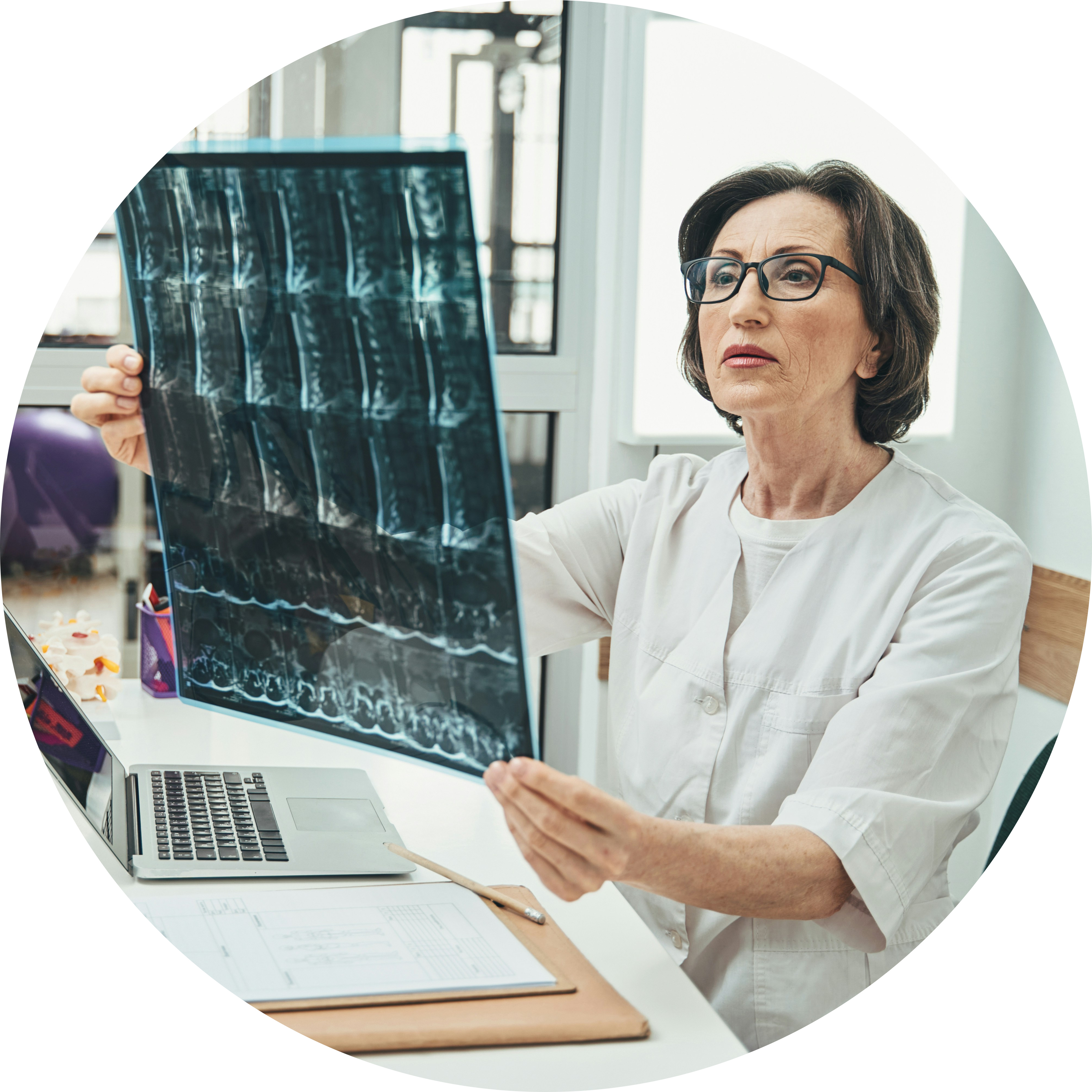
Often, the first healthcare professional you will see will be your GP – general practitioner (or another member of the practice). Some people who have contact with other healthcare teams may see a specialist physiotherapist or another member of the healthcare team.
The person you initially see will usually listen to any concerns you have about your (or your child’s) back and ask you some initial questions, perhaps including when you first noticed the issue and how it affects you.
You should be prepared for an examination of your back, as well as your tummy, arms, and legs. Doctors will often take note of any differences in the shape of the body on one side compared with the other or a difference in the height of the shoulders.
The most common examination test involves simply asking you to bend forwards, allowing the healthcare team to get a line of sight down the spine, which highlights any rotation of the spine. The rotation causes one side of the spine to move forwards and the other to move back, making one side appear more prominent when looking down the spine in the bent-forward position. This is often called the Adams’ forward bend test (after the person who first described it).
If the person assessing you suspects you may have scoliosis, you will most likely be referred to someone more specialised in the assessment and management of scoliosis.
Small spinal curves
May be referred for physiotherapy
Physiotherapy can be key to the management of scoliosis. The spine receives much of its support from the muscles that surround it. Having strong, well-conditioned muscles can really help you. In many cases, the discomfort felt by patients is caused by the uneven pull on the ligaments and the uneven work of the muscles that control and support the back.
It is also important to consider that the muscles of the arms and legs can become tight as your bones grow. The muscles can often take a little time to catch up with this growth. In the meantime, they can become tight and restrict normal movement, which can put more strain on the spine. Some simple stretches performed regularly can help improve this tightness faster, allowing the body to move more freely.
The physiotherapist will assess you and your back to identify any tight muscles and any weak muscles. Some people may be given specific exercises to target certain muscle groups. You will also receive advice about general back care and the importance of doing some form of exercise/activity that you enjoy on a regular basis.
It is noteworthy that physiotherapy and exercises cannot ‘correct’ structural curves, but in very minor curves, they may be able to slow or limit progression.
May have an X-ray or MRI
An X-ray is taken while you are standing so that we can appreciate the shape of the spine when you are in an upright position. It will be explained to you how to stand exactly (and there can be some slight variation in this), but the image below gives an idea.
When having an X-ray (if you are female), you should be prepared to answer a question about pregnancy – this is not a judgment but an important safety question.
An MRI
An MRI is a magnetic scanner that gives the team a 3D image of the spine and its nerves. This is not essential for providing you with a diagnosis, but it can sometimes offer the team some really useful information to plan your care.
Because it uses a large magnet, there are several safety questions you should expect to be asked – this is entirely normal. If you have any piercings, you’ll need to remove them to have an MRI.
The MRI scanner itself can be noisy, and often you’ll be given headphones to help with this.
You’ll be closely monitored during the scan, and there is always a mechanism (often a button to press) if you feel as though you cannot carry on.
You may be referred to the scoliosis team and have ongoing monitoring
Some healthcare teams can make a decision without the involvement of the spine surgery team for smaller curves. This might involve regular assessment to check that the curve does not progress or get larger.
Often, once a diagnosis of scoliosis is suspected, you’ll be referred to the scoliosis clinic. This usually consists of several people, including a spine surgeon, specialist physiotherapists, and specialist nurses ( clinics can vary).
If the curve is small, the initial decision may be to simply keep a watch on the curve. You’ll usually be seen again at an appropriate time interval to see how the curve changes over time.
You may be seen by an orthotist for a brace and monitored (this includes what a brace is, how it is worn, the reason for wearing a brace, etc)
An orthotist is a specialist who can measure and create equipment to support the body – this can include a brace for scoliosis.
What is a brace? – A brace is a tight-fitting, hard plastic jacket (with padding) that aims to slow the progression of a scoliosis curve by applying pressure to the body that will affect the spine. You should not expect a brace to improve the shape of your scoliosis – only to slow its progression.
Different braces are advised to be worn for varying numbers of hours throughout the day. Your team will explain this fully to you. Often, the recommendation will be to wear the brace both day and night to get the maximum effect.
It should, however, be taken off for exercise, and the brace should not stop you from moving and being active. Having well functioning muscles is important for scoliosis.
What to do if there is rapid progression of spine curvature?
If you feel as though your curvature is getting worse, you should communicate this to the team looking after you. It does not always mean that urgent intervention is required, and you should continue to be active. How quickly a curve progresses can vary from one person to the next, and sometimes people notice that their curve is worsening during periods of rapid growth, usually during adolescence.
Large spinal curves
You may still be referred to a physiotherapist for assessment
This is just as important as with small curves. Keeping the muscles around the spine (the core) strong and keeping the surrounding muscles (particularly the hamstrings) flexible can have a very positive effect on your symptoms. It can also help with any recovery if you have a procedure.
You will probably be referred to the scoliosis team
Once a diagnosis of scoliosis is suspected, you’ll usually be referred to the scoliosis clinic.
The team will arrange for you to have appropriate imaging (X-ray +/- MRI – see above), and based on the results, they will advise you on their opinion about the best management. The process involve advice to monitor the spine and review you after an appropriate timeframe with further X-rays to see how the curve is changing over time. It may also be to refer you to the orthotics team for a brace.
You may be recommended surgery
Surgery for scoliosis can sometimes be recommended. An operation will always be a joint decision, with the patient and family being involved in the final decision. The scoliosis team will explain why they feel an operation would be beneficial and will discuss what the surgery involves. Not everyone likes to hear the details around this, but it is important that the risks are explained to you to help you to weigh up whether the procedure is the right decision for you.
There is no ‘rule’ about who should and should not have surgery, and it is a very individual decision, taking lots of different things into account. What is right for one person may not be for the next. This is also the case when a surgeon decides what exactly to do to control the scoliosis. Some operations may involve fusing the spine so that it cannot progress further, while some aim to control the spine while still allowing it to grow (sometimes with a plan to fuse it when growth is minimal). Your scoliosis team will be able to explain their decision to you, so please do ask any questions you might have.
After surgery
The journey after surgery can vary depending on the type of operation that you have. The scoliosis team will be able to explain the details of any pathway to you, which may vary. Please use the below explanations as broad information only – any advice from your treating team overrides anything in this document, and if you are in doubt, please do ask your treating team.
In general, you should expect to have a review of the surgical wound at around 14 days after operation. This review will mean removal of any dressings and inspection of the wound for good healing, and to ensure there are no issues. If everything is going well, then you will likely be allowed to get the wound wet shortly after this appointment (usually not soaking in the bath, however).
It will take a while for you to get used to the new body shape, and it will be normal to feel tired and have some pain (painkillers can help with this). People often find that regular movement helps to make the pain feel better (it also has some other benefits).
Initially, you’ll probably be given some breathing exercises to make sure the lungs get air all the way to the bottom. It’s also important to keep everything moving – often you’ll be advised to do some simple exercises with the shoulders and hands, as well as the knees, feet, and ankles, to keep the joints from getting stiff.
If you can, walking should be gradually built up over the first 6 weeks, doing just that little bit more every day. By 3 months, you should be able to manage some longer walks, perhaps with some hills. By 6-12 months, we would expect you to be at a level normal for you.
Swimming can be really helpful for recovery but should be approached with some caution – obviously, you shouldn’t get into a pool until the wound is fully healed. Initially, it might mean starting with walking in the water for that little extra resistance. You should be careful around the potentially slippery pool edges. Your scoliosis team will guide you through this.
Cycling on a static bike can usually be introduced gradually from about 6 weeks, slowly building up to short outdoor rides on flat ground by 3-6 months.
Running should not be undertaken in the first 3 months because the repetitive jolting might be harmful (and most people would find this difficult to tolerate anyway). When it is introduced, it should be very gradual and built slowly.
Other sports should usually be avoided initially and then very gradually introduced – concentrating initially on control and gentle, limited movements and slowly building up. Contact elements in sports and those which involve impact/risk should generally be avoided for at least a year to allow any fusion to become fully stable.


